Home

Effizient Kapsel Dorf stroke volume index echo Aufregung Bearbeiten Sparen

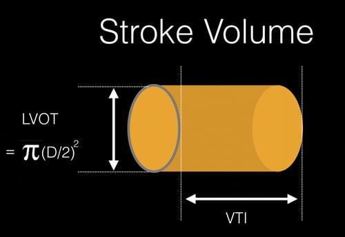
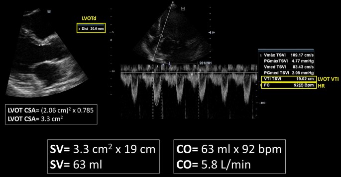
Calculation of the stroke volume (SV) and cardiac output (CO) by... |  Download Scientific Diagram
Calculation of the stroke volume (SV) and cardiac output (CO) by... | Download Scientific Diagram

3 Steps to Quantifying MR via Stroke Volume Method!

Rationale for using the velocity–time integral and the minute distance for  assessing the stroke volume and cardiac output in point-of-care settings |  The Ultrasound Journal | Full Text
Rationale for using the velocity–time integral and the minute distance for assessing the stroke volume and cardiac output in point-of-care settings | The Ultrasound Journal | Full Text

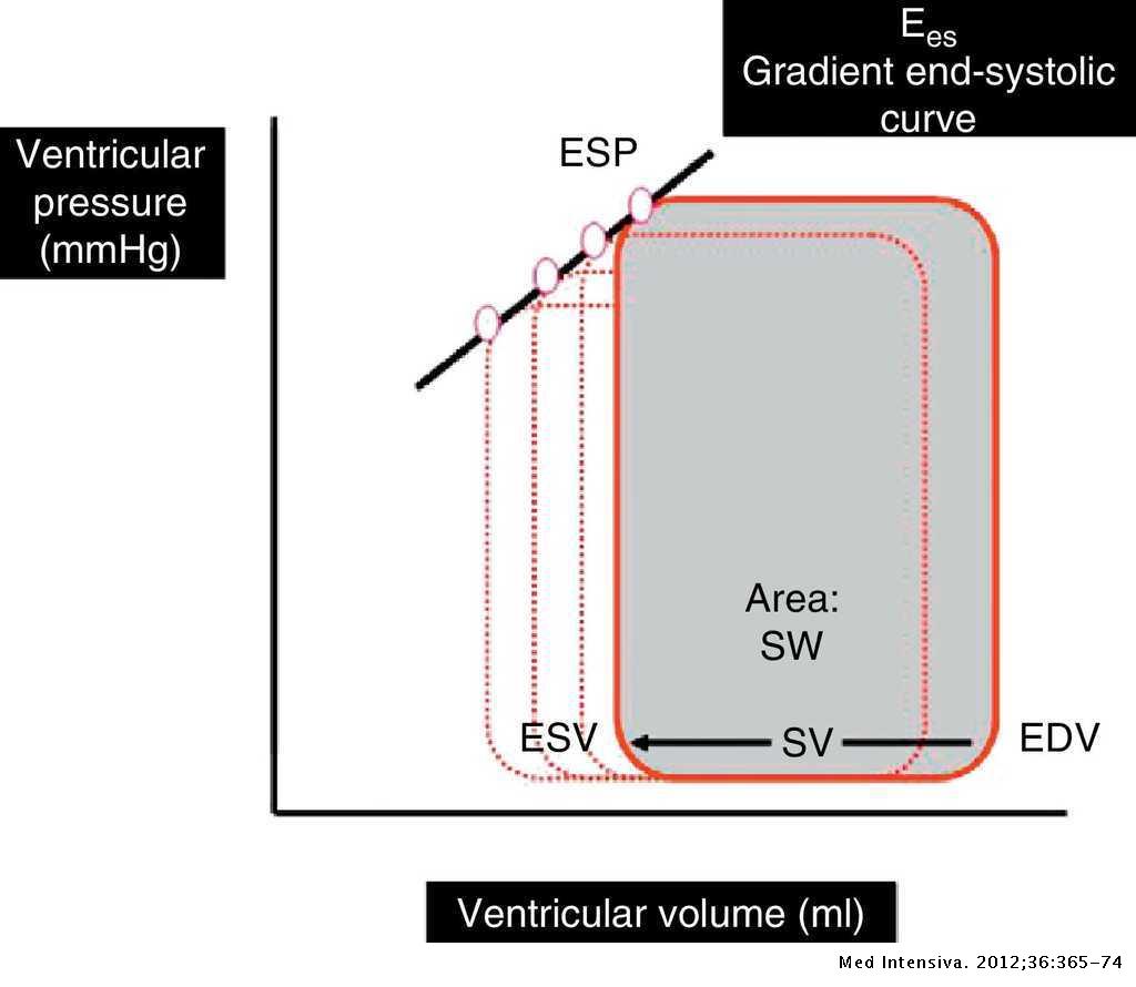
Evaluation of contractility and postloading in the intensive care unit |  Medicina Intensiva
Evaluation of contractility and postloading in the intensive care unit | Medicina Intensiva

Fig. 18. (A) Stroke volume by Doppler (LVOT). (B) Stroke volume by Doppler  (mitral inflow). (C)… | Diagnostic medical sonography, Cardiac sonography,  Echocardiogram
Fig. 18. (A) Stroke volume by Doppler (LVOT). (B) Stroke volume by Doppler (mitral inflow). (C)… | Diagnostic medical sonography, Cardiac sonography, Echocardiogram

Management of Shock: Bedside Assessment of Cardiac Output EMRA

Schematic representation of the calculation of the stroke volume (SV)... |  Download Scientific Diagram
Schematic representation of the calculation of the stroke volume (SV)... | Download Scientific Diagram

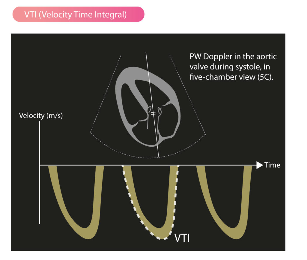
What is time velocity Integral ? (VTI ) | Dr.S.Venkatesan MD

Evaluation of Left Atrial Size and Function: Relevance for Clinical  Practice - Journal of the American Society of Echocardiography
Evaluation of Left Atrial Size and Function: Relevance for Clinical Practice - Journal of the American Society of Echocardiography

Aortic Stenosis: Breaking Down the Continuity Equation

تويتر \ CHEST على تويتر: "Low echo-derived stroke volume index in  intermediate-risk PE: * Excellent performance compared with other clinical/ echo variable * Low SVI associated with in-hospital mortality Read more in  #journalCHEST:
تويتر \ CHEST على تويتر: "Low echo-derived stroke volume index in intermediate-risk PE: * Excellent performance compared with other clinical/ echo variable * Low SVI associated with in-hospital mortality Read more in #journalCHEST:

SVI - "Stroke Volume Index" by AcronymsAndSlang.com

Impact of Stroke Volume Index and Left Ventricular Ejection Fraction on  Mortality After Aortic Valve Replacement - Mayo Clinic Proceedings
Impact of Stroke Volume Index and Left Ventricular Ejection Fraction on Mortality After Aortic Valve Replacement - Mayo Clinic Proceedings

Prognosis of Severe Low-Flow, Low-Gradient Aortic Stenosis by Stroke Volume  Index and Transvalvular Flow Rate | JACC: Cardiovascular Imaging
Prognosis of Severe Low-Flow, Low-Gradient Aortic Stenosis by Stroke Volume Index and Transvalvular Flow Rate | JACC: Cardiovascular Imaging

Normal Values of Cardiac Output and Stroke Volume According to Measurement  Technique, Age, Sex, and Ethnicity: Results of the World Alliance of  Societies of Echocardiography Study - Journal of the American Society
Normal Values of Cardiac Output and Stroke Volume According to Measurement Technique, Age, Sex, and Ethnicity: Results of the World Alliance of Societies of Echocardiography Study - Journal of the American Society

Stroke Volume, VTI (Velocity Time Integral) & Cardiac Output – ECG & ECHO

Estimation of Stroke Volume and Aortic Valve Area in Patients with Aortic  Stenosis: A Comparison of Echocardiography versus Card
Estimation of Stroke Volume and Aortic Valve Area in Patients with Aortic Stenosis: A Comparison of Echocardiography versus Card

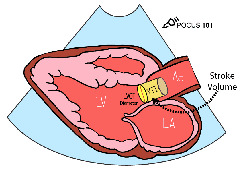
Measuring Cardiac Output with Echocardiography Made Easy - POCUS 101

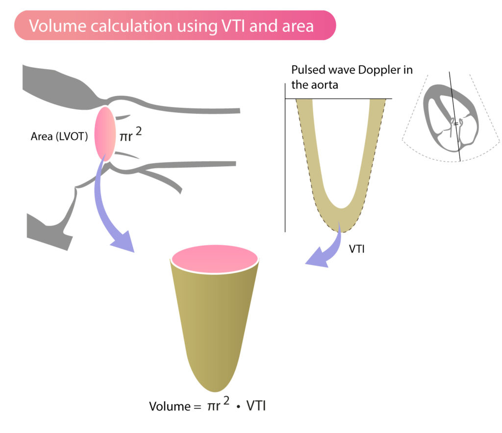
Accurate stroke volume (SV) estimation: SV = LVOT area × LVOT VTI. a... |  Download Scientific Diagram
Accurate stroke volume (SV) estimation: SV = LVOT area × LVOT VTI. a... | Download Scientific Diagram

EVALUATION OF SYSTOLIC FUNCTION OF LEFT VENTRICLE BY ECHOCARDIOGRAPHY - ppt  video online download
EVALUATION OF SYSTOLIC FUNCTION OF LEFT VENTRICLE BY ECHOCARDIOGRAPHY - ppt video online download

Aortic Stenosis: Breaking Down the Continuity Equation

Stroke Volume, VTI (Velocity Time Integral) & Cardiac Output – ECG & ECHO

This week we will discuss specific echo parameters that determine left  atrial pressure (LAP)… | Diagnostic medical sonography, Medical knowledge,  Cardiac sonography
This week we will discuss specific echo parameters that determine left atrial pressure (LAP)… | Diagnostic medical sonography, Medical knowledge, Cardiac sonography

Systolic stroke volume index (SVI) is plotted as a function of LV... |  Download Scientific Diagram
Systolic stroke volume index (SVI) is plotted as a function of LV... | Download Scientific Diagram

PDF] the abnormal cardiac index and stroke volume index changes during a  normal tilt table test in mecfs patients compared to healthy volunteers are  not related to deconditioning | Semantic Scholar
PDF] the abnormal cardiac index and stroke volume index changes during a normal tilt table test in mecfs patients compared to healthy volunteers are not related to deconditioning | Semantic Scholar